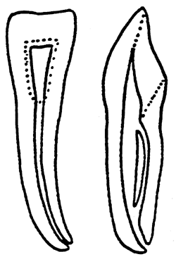

The canines (tooth C, H) have a trapezoidal crown with one labial cusp. Concavoconvex in shape, their labial surface is convex, and their palatal surface is concave.Ĭanines – The primary teeth canines are also morphologically similar to permanent teeth canines. Incisors are generally single-rooted and also have one root canal. They also prominently figure into the aesthetic of the oral region. The central incisor is larger than the lateral incisor, and the maxillary central incisor is the largest of all the incisors. They consist of the central incisors(tooth E, J) and the lateral incisors (tooth D, G). Incisors – The primary teeth incisors are essentially smaller morphological versions of permanent teeth incisors. The incisors are used for cutting food and are therefore sharp-edged in shape. The two incisors constituting the central and lateral incisors. The primary teeth are grouped into four quadrants, each containing two incisors, a canine, and two molars. The alveolar bone supports the root of the tooth and contains sockets that contain the embedded roots of teeth. The tip of each root contains a tiny opening called an apical foramen, where external blood vessels and nerves enter the root. The accessory canals supply blood vessels and nerves to the pulp and arise from the main root canal. In addition to their stabilizing role, they also serve as shock absorbers. The periodontal ligament is composed of connective tissue fibers that anchor the tooth to the alveolar bone. Pulp allows for the tooth's ability to feel temperature, touch, and pain via stimulation of dentin fibrils. Deep to the dentin is the dental pulp which is composed of innervated, vascular tissue. Reparation of dentin occurs with the production of secondary dentin. Dentin is composed of hollow tubules that contain fibrils and contain sensory endings. Dentin is living tissue and is enriched by dentinal tubules that run from the pulp. Normally lying beneath the gingiva, the cementum is composed of calcium hydroxyapatite and allows for attachment to the periodontal ligament.Ī layer of innervated, porous tissue called dentin lies beneath the enamel and cementum and constitutes the bulk of the tooth. Enamel can form fluorapatite crystals when exposed to fluoride to become harder and more resistant to caries.Ĭementum is the softer outer tissue surrounding the root that can be damaged by periodontal disease. Owing to their avascularity, they remineralize but do not grow after an acid attack. Enamel is primarily composed of hydroxyapatite and contains no nerves or blood vessels. Enamel color is determined by enamel thickness. Enamel is an avascular, hard material that protects the outer tooth, gives the tooth its whitish color, and is first affected by dental caries.

The anatomic crown is covered by enamel, while the anatomic root is covered by cementum. The tooth is divided into the crown and the root. Primary teeth are labeled in the following classification system and are designated in a clockwise direction:Įruption(appearance) and exfoliation(shedding) of the primary dentition occur in the following order: Since primary teeth are shed, as some trees shed their leaves, they are also referred to as deciduous teeth, which are eventually replaced by the adult permanent dentition. Incisors and canines can be classified further as "anterior teeth" and molars and premolars as "posterior teeth." The primary teeth erupt during the first year and into the third years of life and are then exfoliated(shed) between the ages of 6 and 12. When divided on the mid-line sagittal plane, these teeth are organized into the right and left halves. The primary teeth are organized in two arches: the maxillary (upper) arch and the mandibular (lower) arch. The primary dentition constitutes the first teeth to erupt in the pediatric patient. Comprised of 20 teeth, they are labeled based on an alphabetical system rather than the numbering system used for permanent teeth.


 0 kommentar(er)
